Engineered PSF for High-NA Microscopy Imaging

The microscopy imaging techniques are developed rapidly in recent decades. The PSF (Point Spread Function) is very often not an Airy disk at the image plane. A donut shape can be engineered when imaging a dipole source orientated along the longitudinal axis. We demonstrate in VirtualLab Fusion that different asymmetric PSFs, which are not Airy disks, are obtained when the orientation of the dipole source changes. Moreover, a double-helix PSF can be obtained by inserting a certain phase mask in the pupil plane of a microscopy system [Ginni Grover et al., Opt. Exp. 2012]. With such an engineered PSF, even a small defocus of the object can be observed, i.e., the axial resolution can be improved drastically compared to the traditional imaging approach. We demonstrate this phenomenon by applying a commercial microscopy lens (Nikon) system in VirtualLab Fusion.

Analysis of PSF of a Dipole Source by a High-NA Microscopy System

In VirtualLab Fusion, the PSF of a dipole source can be analyzed in a straightforward manner. It is demonstrated that the PSFs have different shapes when the orientation of the dipole source changes.

Double-Helix PSF for 3D Imaging Microscopy

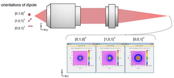
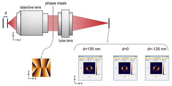
In VirtualLab Fusion, the double-helix PSF be analyzed in a straightforward manner by inserting a phase mask in the pupil plane of a high-NA microscopy system. It is demonstrated that the double-helix PSF rotates tangibly even when there is only a small defocus (~130 nm) of the object point.

Register For Our Next Webinar


Register For Our Next Webinar